Home
/ Chest Muscles Diagram - If You Only Train Your Chest Muscle You Ll End Up Looking Worse, Superficial muscles are the muscles closest to the skin surface and can usually be seen while a body is performing actions.
Chest Muscles Diagram - If You Only Train Your Chest Muscle You Ll End Up Looking Worse, Superficial muscles are the muscles closest to the skin surface and can usually be seen while a body is performing actions.
Chest Muscles Diagram - If You Only Train Your Chest Muscle You Ll End Up Looking Worse, Superficial muscles are the muscles closest to the skin surface and can usually be seen while a body is performing actions.. The word sternum originates from the ancient greek word 'sternon', meaning chest. It contains organs including the heart lungs and thymus gland as well as muscles and various other internal structures. The major muscle in the chest is the pectoralis major. Chest muscle in women body, find out more about chest muscle in women body. Human anatomy for muscle, reproductive, and skeleton.
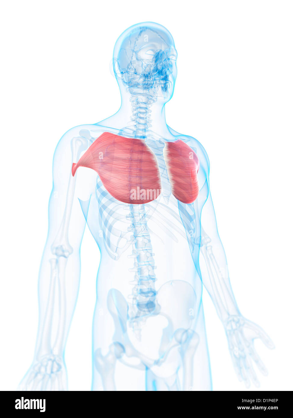
The sternum is also known as the breastbone. Of the two chest muscles, the pectoralis major (a.k.a. The chest muscles the 2 pectoralis muscles, the pectoralis major and the pectoralis minor (the larger and smaller muscles of the chest) connect the front of the chest wall with the humerus (upper arm bone) and shoulder (fig). Related posts of anatomy of the chest and stomach abdominal regions and organs. The chest muscles are made up of the pectoralis major and, underneath that, the pectoralis minor.
Barbell Bench Press Exercise Guide Traineatgain Com from d3p2750kqzf48b.cloudfront.net It is a flat bone that articulates with the clavicle and the costal cartilages of the upper 7 ribs (true ribs), while the 8th, 9th and 10th ribs (false ribs) are indirectly attached with sternum via costal cartilage of the ribs above. The crossword solver found 20 answers to the chest muscle crossword clue. The pec major) is the one that commands the most real estate. Back muscles anatomy video 12 photos of the back muscles anatomy video back muscles anatomy images, back muscles anatomy video, human muscles, back muscles anatomy images, back muscles anatomy video. Related posts of chest muscle diagram muscle gross anatomy. There are red muscles stretched over the stomach, chest, and shoulders, and on top of each breast is a complicated structure made out of milk ducts, which appears in pieces fanned out that make it. The primary function is certainly to provide support to the skeletal system and to facilitate its movements. Chest muscle in women body, find out more about chest muscle in women body.
Enter the answer length or the answer pattern to get better results.
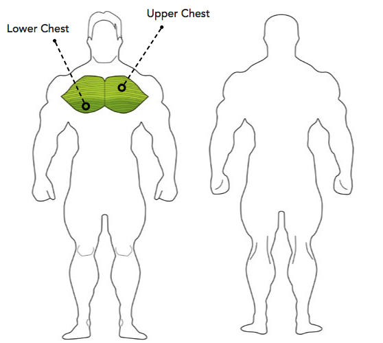
Related posts of muscle diagram for chest and back back muscles anatomy video. Many in the neck help to stabilize or move the head. The usual cause of overstretched chest muscles can be the over exercising. The dominant muscle in the upper chest is the pectoralis major. Chest muscles, chest muscle diagram. There are multiple functions of these chest muscles. Several muscles that move the arms, head, and neck have their origins on the sternum. Muscle gross anatomy 12 photos of the muscle gross anatomy gross anatomy of cardiac muscle, gross anatomy of skeletal muscle worksheet, gross muscle anatomy test, muscle gross anatomy quiz, muscular system gross anatomy chapter 10, human muscles, gross anatomy of cardiac muscle, gross anatomy of skeletal muscle worksheet, gross. The chest, as part of this group, enables you to perform pushing actions such as the barbell bench press or a daily activity such as moving a heavy dresser. Chest wall pain is caused by problems affecting the muscles, bones and/or nerves of the chest wall. Human body muscle system, the muscles of the human body that work the skeletal system, that are under voluntary control, and that are concerned with movement, posture, and balance. It also protects several vital organs of the chest, such as the heart, aorta, vena cava, and thymus gland that are located just deep to the sternum. To fully develop your chest, you need to hit it with heavy weight using a couple smartly chosen exercises.
Create bulk of the chest, commonly known as pecs sternoicleidomastoid connects from the mastoid process at the back/base of head to the sternum and clavicle Abdominal regions and organs 12 photos of the abdominal regions and organs 9 abdominal regions and its organs, abdominal cavity regions and organs, abdominal regions and associated organs, abdominal regions and its organs, abdominal regions and quadrants and organs, human anatomy, 9 abdominal regions and its. Human anatomy for muscle, reproductive, and skeleton. It contains organs including the heart lungs and thymus gland as well as muscles and various other internal structures. Many in the neck help to stabilize or move the head.
Thoracic And Abdominal Muscles Lecturio Online Medical Library from philschatz.com Pain, which may be sharp (an acute pull) or dull (a chronic strain) swelling. The sternum is also known as the breastbone. Doctors diagnose chest wall pain in at least 25% of patients who come to the emergency room for chest pain. It is a flat bone that articulates with the clavicle and the costal cartilages of the upper 7 ribs (true ribs), while the 8th, 9th and 10th ribs (false ribs) are indirectly attached with sternum via costal cartilage of the ribs above. Several muscles that move the arms, head, and neck have their origins on the sternum. Muscle gross anatomy 12 photos of the muscle gross anatomy gross anatomy of cardiac muscle, gross anatomy of skeletal muscle worksheet, gross muscle anatomy test, muscle gross anatomy quiz, muscular system gross anatomy chapter 10, human muscles, gross anatomy of cardiac muscle, gross anatomy of skeletal muscle worksheet, gross. Click the answer to find similar crossword clues. The pectoral region is located on the anterior chest wall.
The chest muscles the 2 pectoralis muscles, the pectoralis major and the pectoralis minor (the larger and smaller muscles of the chest) connect the front of the chest wall with the humerus (upper arm bone) and shoulder (fig).
Several muscles that move the arms, head, and neck have their origins on the sternum. Each one spans half of the upper chest, and has attachment points on the sternum (breastbone), ribs, clavicle (collarbone), and humerus. The chest muscles of our body can get pulled and strained. It also protects several vital organs of the chest, such as the heart, aorta, vena cava, and thymus gland that are located just deep to the sternum. The word sternum originates from the ancient greek word 'sternon', meaning chest. In this image, you will find frontalis, orbicularis oculi, zygomaticus, masseter, orbicularis oris, sternocleidomasteoid, deltoid, pectoralis major, biceps brachii, iliopsoas, adductor longus, gastrocnemius. Function of the chest muscles. The pectoralis major, pectoralis minor, serratus anterior and subclavius. Chest muscles, chest muscle diagram. Difficulty moving the affected area. To fully develop your chest, you need to hit it with heavy weight using a couple smartly chosen exercises. The chest muscles are made up of the pectoralis major and, underneath that, the pectoralis minor. Related posts of chest muscle diagram muscle gross anatomy.
Chest wall pain is caused by problems affecting the muscles, bones and/or nerves of the chest wall. Below is a diagram showing the chest muscles depicting where the different exercises target. The chest, as part of this group, enables you to perform pushing actions such as the barbell bench press or a daily activity such as moving a heavy dresser. Classic symptoms of strain in the chest muscle include: The usual cause of overstretched chest muscles can be the over exercising.
Human Chest Muscles High Resolution Stock Photography And Images Alamy from c8.alamy.com To fully develop your chest, you need to hit it with heavy weight using a couple smartly chosen exercises. Chest muscles, chest muscle diagram. The chest muscles are made up of the pectoralis major and, underneath that, the pectoralis minor. Abdominal regions and organs 12 photos of the abdominal regions and organs 9 abdominal regions and its organs, abdominal cavity regions and organs, abdominal regions and associated organs, abdominal regions and its organs, abdominal regions and quadrants and organs, human anatomy, 9 abdominal regions and its. The chest muscles the 2 pectoralis muscles, the pectoralis major and the pectoralis minor (the larger and smaller muscles of the chest) connect the front of the chest wall with the humerus (upper arm bone) and shoulder (fig). The chest is part of a larger group of pushing muscles found in the upper body. It contains four muscles that exert a force on the upper limb: Create bulk of the chest, commonly known as pecs sternoicleidomastoid connects from the mastoid process at the back/base of head to the sternum and clavicle
In this image, you will find frontalis, orbicularis oculi, zygomaticus, masseter, orbicularis oris, sternocleidomasteoid, deltoid, pectoralis major, biceps brachii, iliopsoas, adductor longus, gastrocnemius.
The word sternum originates from the ancient greek word 'sternon', meaning chest. Related posts of chest muscle diagram muscle gross anatomy. Chest muscle in women body, find out more about chest muscle in women body. I often get asked, how can i build thick powerful pecs? The usual cause of overstretched chest muscles can be the over exercising. The chest muscles of our body can get pulled and strained. The dominant muscle in the upper chest is the pectoralis major. Chest wall pain is caused by problems affecting the muscles, bones and/or nerves of the chest wall. Related posts of anatomy of the chest and stomach abdominal regions and organs. The chest muscles are made up of the pectoralis major and, underneath that, the pectoralis minor. The heart is the epicenter of the. Human anatomy for muscle, reproductive, and skeleton. Each one spans half of the upper chest, and has attachment points on the sternum (breastbone), ribs, clavicle (collarbone), and humerus.
